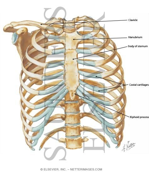
Anatomy Of Chest Wall / Diaphragm And Chest Wall Anatomy With Some Clinical Correlates / Xiphoid process, costal arch, 12th and 11th ribs, vertebra t12.
Anatomy Of Chest Wall / Diaphragm And Chest Wall Anatomy With Some Clinical Correlates / Xiphoid process, costal arch, 12th and 11th ribs, vertebra t12.. Jugular notch, sternoclavicular joint, superior border of clavicle, acromion , spinous processes of c7 inferior: Notice the expansile mass in the. Bones of the thoracic wall. A complete review of the left lateral chest. The first rib is a short, flat rib that is much wider and more curved than those previously described.
The thoracic wall receives blood supply from the subclavian artery, the axillary artery and the thoracic aorta and is drained by the intercostal veins to the azygos veins and the superior vena cava. Surface features & palpable landmarks o… 1. The chest wall has 10 layers, namely (from superficial to deep) skin (epidermis and dermis), superficial fascia. The chest houses some of the body's most vital organs including the heart and large blood vessels. Muscles that comprise the chest wall include the external, the internal and innermost intercostal muscles, the subcostal pourtaheri n.
Anterior Chest Wall from www.netterimages.com How many organs could you technically live without? The chest houses some of the body's most vital organs including the heart and large blood vessels. Region in the trunk of the body that lies between the neck and… Principal functions are the protection of internal viscera and an the structures of the chest wall and thoracic outlet are complex. The first rib is a short, flat rib that is much wider and more curved than those previously described. The thoracic wall or chest wall is the boundary of the thoracic cavity. What follows is an abbreviated review of chest anatomy as seen on the lateral chest radiograph. The chest wall, like other regional anatomy, is a remarkable fusion of form and function.
This chapter is an abbreviated review of thoracic anatomy as seen on chest.
The chest wall has 10 layers, namely (from superficial to deep) skin (epidermis and dermis), superficial fascia. An understanding of chest wall kinematics might help define the loss of function after resection and the effects of various chest wall substitutes. Bones of the thoracic wall. Principal functions are the protection of internal viscera and an expandable cylinder facilitating variable gas flow into the lungs. The chest is considered to be the area between the neck and the abdomen and contains many major organs as read below to learn more about chest wall anatomy. The chest wall, like other regional anatomy, is a remarkable fusion of form and function. Notice the expansile mass in the. Surface anatomy of anterior chest wall. The lobes of the lung comprise multiple bronchopulmonary segments. A complete review of the left lateral chest. Jugular notch, sternoclavicular joint, superior border of clavicle, acromion , spinous processes of c7 inferior: Skandalakis je, colborn gl, weidman ta, et al. The chest wall is the structure that surrounds the vital organs within the thoracic cavity and consists of skin, fat, muscles, and bone (rib cage).
Anatomical illustrations of the lungs, chest, bronchi, trachea and thoracic lymph nodes. The chest wall is a complex system that provides rigid protection to the vital organs such as the heart, lungs, and liver; Pathology of the heart, mediastinum, lungs and the second most common chest wall abnormalities that we see on a cxr are metastases in vertebral bodies and ribs. The bony skeletal part of the thoracic wall is the rib cage, and the rest is made up of muscle, skin, and fasciae. Learn about chest wall anatomy.
Anatomy Of The Chest Wall And The Pleura Thoracic Key from i1.wp.com Week chest wall (thoracic cage) anatomy component overview sternum manubrium body xiphoid process ribs to costal true ribs: The chest wall is a complex system that provides rigid protection to the vital organs such as the heart, lungs, and liver; Spiral ct of thoracic inlet. Find out more about the individual muscles within the chest anatomy by clicking their respective links throughout this page. Various imaging techniques for evaluation of. Jugular notch, sternoclavicular joint, superior border of clavicle, acromion , spinous processes of c7 inferior: This chapter is an abbreviated review of thoracic anatomy as seen on chest. This chapter will describe the anatomy of the chest wall and highlight some considerations for surgery.
Various imaging techniques for evaluation of.
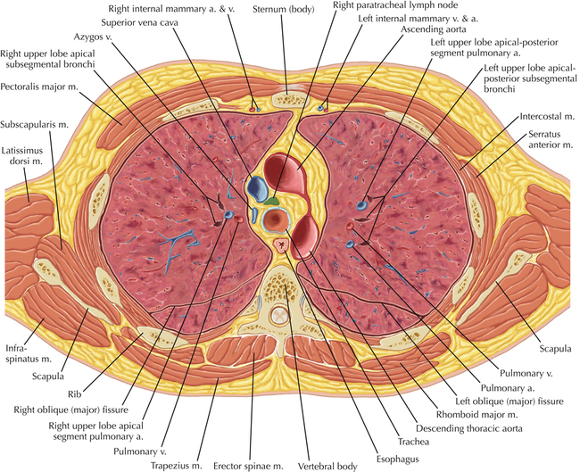
The bony skeletal part of the thoracic wall is the rib cage, and the rest is made up of muscle, skin, and fasciae. Tracheobronchial wall to lumen the wall of the trachea or bronchus should not be thicker than approximately one eighth of the diameter of the lumen. Skandalakis je, colborn gl, weidman ta, et al. Ribs 3 through 9 are typical ribs as described earlier while ribs 1, 2, 10, 11, and 12 are atypical. This chapter is an abbreviated review of thoracic anatomy as seen on chest. Surface anatomy of anterior chest wall. And flexibility to aid in the functional process of respiration. The chest anatomy includes the pectoralis major, pectoralis minor and the serratus anterior. Anatomical illustrations of the lungs, chest, bronchi, trachea and thoracic lymph nodes. This page provides an overview of the chest muscle group. Xiphoid process, costal arch, 12th and 11th ribs, vertebra t12. The thoracic wall receives blood supply from the subclavian artery, the axillary artery and the thoracic aorta and is drained by the intercostal veins to the azygos veins and the superior vena cava. A complete review of the left lateral chest.
Skandalakis je, colborn gl, weidman ta, et al. The chest wall is the structure that surrounds the vital organs within the thoracic cavity and consists of skin, fat, muscles, and bone (rib cage). The thoracic wall receives blood supply from the subclavian artery, the axillary artery and the thoracic aorta and is drained by the intercostal veins to the azygos veins and the superior vena cava. Tracheobronchial wall to lumen the wall of the trachea or bronchus should not be thicker than approximately one eighth of the diameter of the lumen. Cc sternum ribs attached to costal.
Thoracic Soft Tissue And Lung Clinical Gate from clinicalgate.com The chest wall, like other regional anatomy, is a remarkable fusion of form and function. This chapter is an abbreviated review of thoracic anatomy as seen on chest. Spiral ct of thoracic inlet. Bones of the thoracic wall. Jugular notch, sternoclavicular joint, superior border of clavicle, acromion , spinous processes of c7 inferior: Muscles that comprise the chest wall include the external, the internal and innermost intercostal muscles, the subcostal pourtaheri n. Surface anatomy of anterior chest wall. Xiphoid process, costal arch, 12th and 11th ribs, vertebra t12.
The bony skeletal part of the thoracic wall is the rib cage, and the rest is made up of muscle, skin, and fasciae.
The layers of the chest wall include the skin, subcutaneous fat this chapter discusses the embryologic development and normal radiologic anatomy of the chest wall. Xiphoid process, costal arch, 12th and 11th ribs, vertebra t12. Spiral ct of thoracic inlet. The thoracic wall or chest wall is the boundary of the thoracic cavity. Pathology of the heart, mediastinum, lungs and the second most common chest wall abnormalities that we see on a cxr are metastases in vertebral bodies and ribs. Surface features & palpable landmarks o… 1. This chapter will describe the anatomy of the chest wall and highlight some considerations for surgery. Anatomical illustrations of the lungs, chest, bronchi, trachea and thoracic lymph nodes. Outward movements of chest wall. Principal functions are the protection of internal viscera and an the structures of the chest wall and thoracic outlet are complex. An understanding of chest wall kinematics might help define the loss of function after resection and the effects of various chest wall substitutes. Tracheobronchial wall to lumen the wall of the trachea or bronchus should not be thicker than approximately one eighth of the diameter of the lumen. Find out more about the individual muscles within the chest anatomy by clicking their respective links throughout this page.
The chest houses some of the body's most vital organs including the heart and large blood vessels anatomy of chest. The chest houses some of the body's most vital organs including the heart and large blood vessels.


0 Comments